Hip Muscles
 Anatomy & Physiology
Introduction & Overview
There are a number of ways that the muscles of the hips are categorized and described, including the following:
- Location — The hip muscles include those on all four sides of the hip joint: Anterior, Medial, Posterior, and Lateral.
- Function — When categorized by function, the hip muscles are described as: Abductors, Adductors, Flexors, Extensors and Rotators.
- Muscle Group — Another way they may be categorized is by muscle group: Quadriceps, Hamstrings, Adductors and Gluteals.
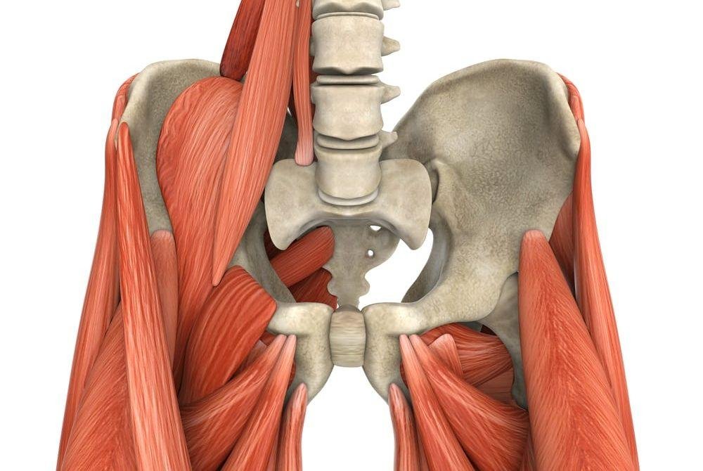
MUSCLES PULL THE FEMUR IN MANY DIRECTIONS
[Because the hip] joint has the possibility to move at so many different angles, you must have muscles that can pull the femur in all of these directions, too. The tissues around the hip joint do just that; they attach to and cross the hip joint at all of the angles necessary to move it effectively. It goes without saying that any of the muscles, if tight, can also limit the hip’s range of motion. – David Keil
Anterior Muscles

Overview
- The anterior muscles are on the front of the body and include the hip flexors and knee extensors.
- They contract when sitting up or lifting a leg to step up.
- Anterior muscles include the hip flexors, quadratus lumborum and quadriceps.
Hip Flexors
- Muscles: Psoas, Illiacus, Rectus femoris (the only hip flexor to cross the hip and knee and can have actions at both); the iliopsoas is comprised of the psoas major and iliacus muscles
- Description: These muscles connect the front of the pelvis to the front of the thigh.
Teaching Considerations
- Hip flexors can be a limiting factor in backbends, inhibiting the pelvis from rotating backward over the femurs.
Quadriceps
- Muscles: Rectus Femoris, Vastus Lateralis, Vastus Medialis, Vastus Intermedius
- Description: These muscles originate at the top of the femur (thigh bone) and attach to the front of the tibia (shinbone).
- Actions: The quadricep muscles extend the leg at the knee joint.
Teaching Considerations
- Quads are strengthened by standing poses.
- They are stretched by lunges and backbends.
Medial Muscles (Adductors)
Overview
- The medial muscles run down the inside of the leg and are called the adductors.
- Adductors move the thigh toward the body’s midline.
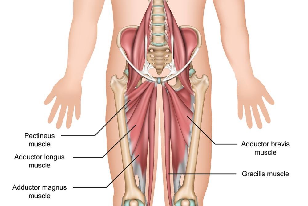
Adductors
- Muscles: Pectineus, Adductor Longus, Adductor Brevis, Adductor Magnus, Gracilis
- Description: The five adductors run from the pelvis down the inside of the leg.
- Actions: These muscles are responsible for adducting the thigh at the hip joint and flexing the thigh at the hip joint.
Teaching Considerations
- The adductors can limit poses that require the thighs to separate widely such as in Baddha Konasana and Upavistha Konasana.
FASCIAL CONNECTIONS
[The adductors are] the top end of a fascial line that begins at the arch of the foot and runs up the inside line of the leg ending at the pelvic floor. [Collapsed arches, pronation of the feet or standing with feet and hips] ducked out in a ballerina kind of position cause this inner foundation to sag, and weakens the whole pelvic girdle. – Robin Rothenberg
Posterior Muscles
Overview
- The posterior muscles are on the back body and include the hamstrings and glutes.
- These muscles straighten the thigh.
- Hamstrings contract when standing, walking, running and climbing stairs.
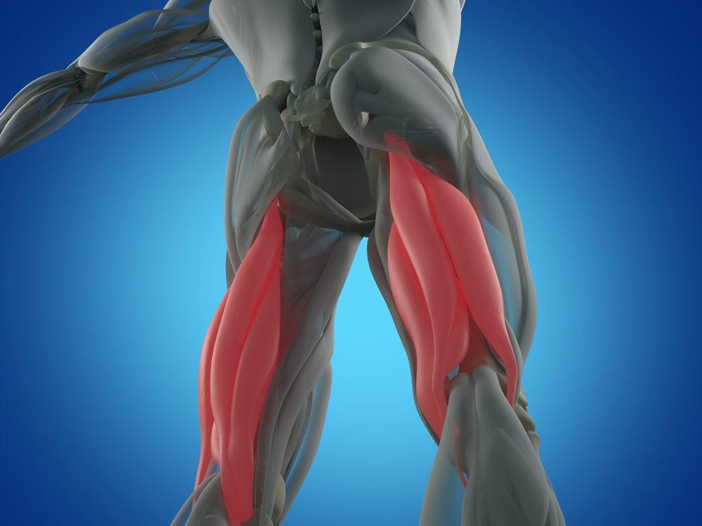
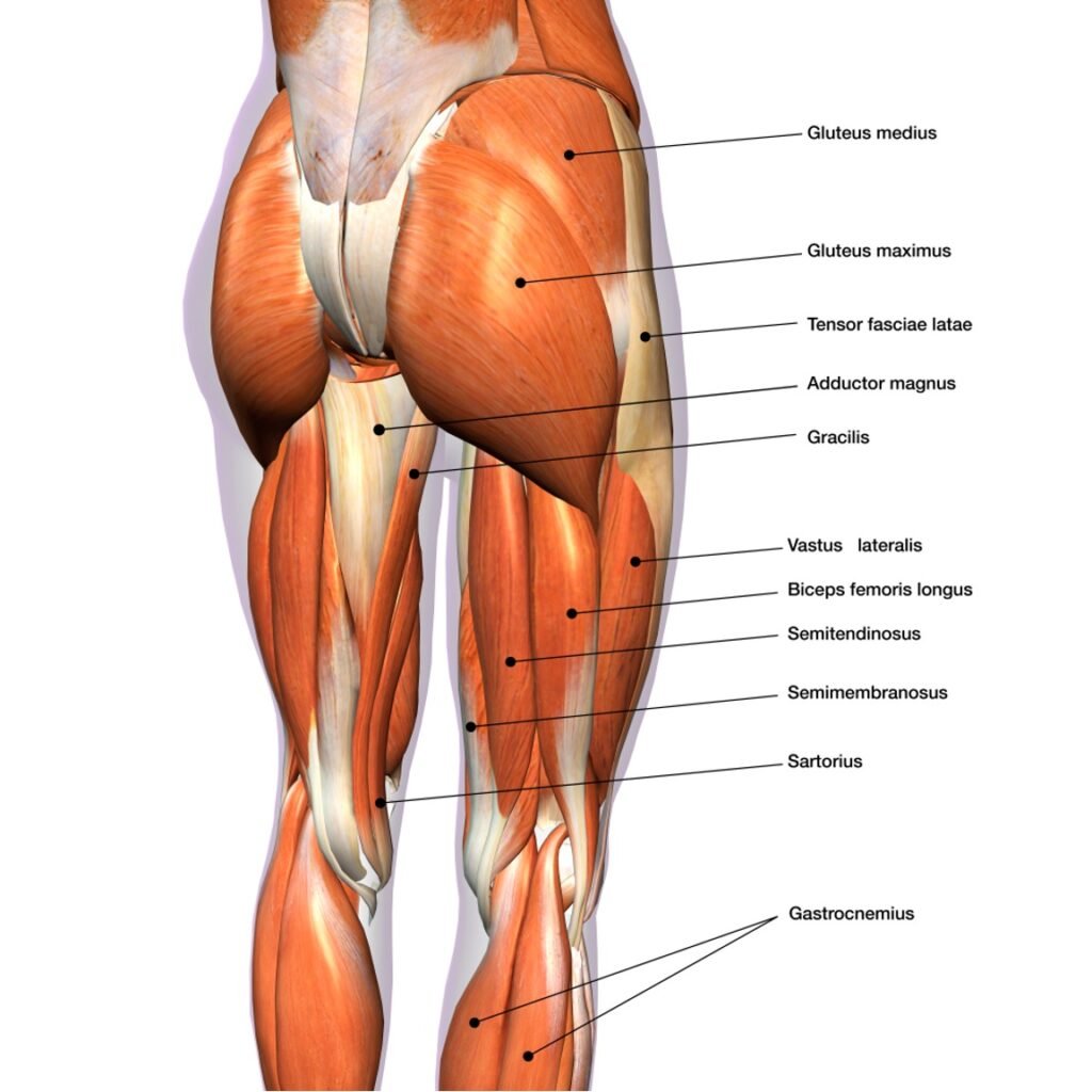
Hamstrings
- Muscles: Biceps Femoris, Semitendinosus, Semimembranosus
- Description: The hamstrings attach to the ischial tuberosity at the back of the pelvis and run down the back of the leg.
- Actions: These muscles flex the leg at the knee joint, extend the thigh at the hip joint, and medially rotate the leg at the knee joint.
Teaching Considerations
- When tight, these muscles can pull and stress the low back.
- The hamstrings can limit forward bending because they keep the pelvis from rotating forward.
- These muscles are stretched by hip flexion, including forward bends such as Paschimottanasana in which the knee is straightened while the hip joint flexes.
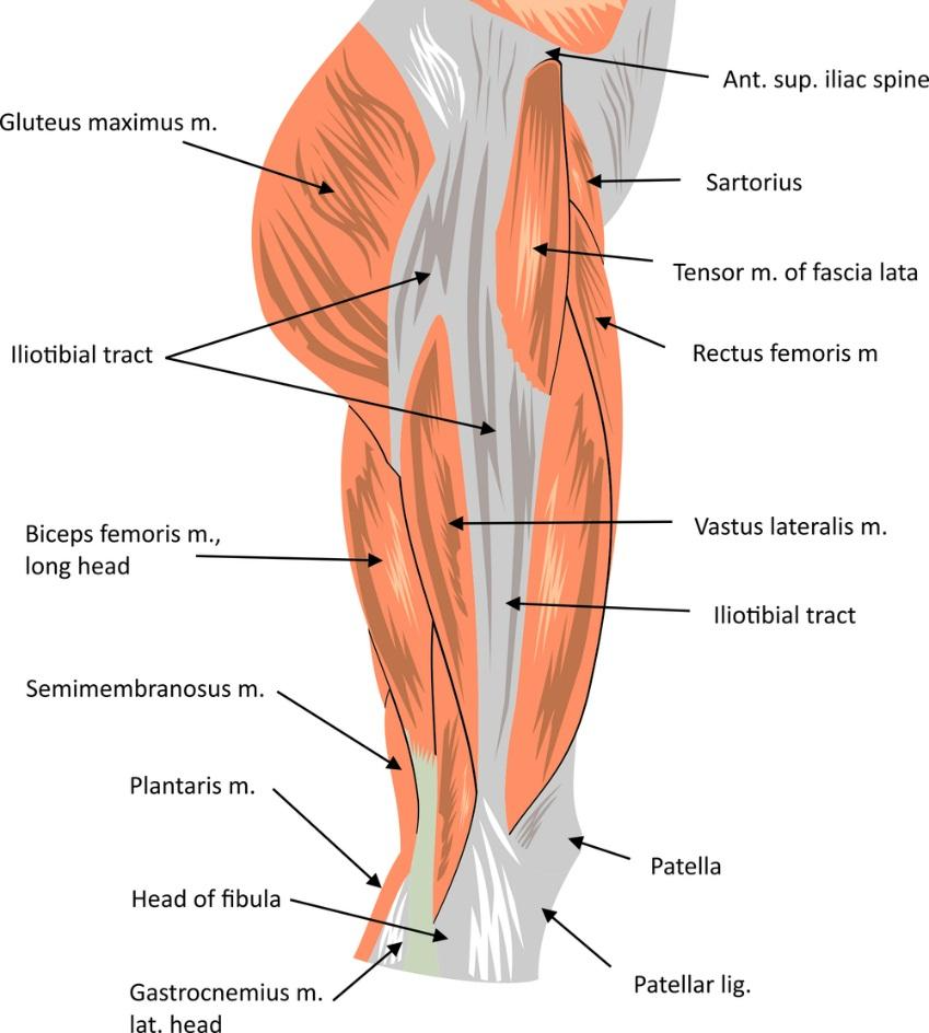
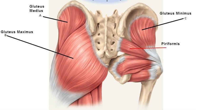
Abductors / Gluteus Muscles
- Muscles: Gluteus Maximus (the largest, most posterior and most superficial), Gluteus Medius (medium-sized, fan-shaped partially covering the glute maximus and covering the glute minimus), Gluteus Minimus
- Description: These muscles run from the outside of the pelvis to the outside of the femur; they work opposite the adductors. The gluteus maximus externally rotates the hip and assists in knee extension. It attaches along the outer portion of the femur and connects through fascia to the iliotibial (IT) band of the tensor fascia lata (TFL).
- Actions: The abductors are responsible for lateral rotation of the thigh at the hip joint, extension of the thigh at the hip joint, and abduction of the thigh at the hip joint.
Teaching Considerations
- Tightness in the glute maximus limits forward bending at the hips.
- Abductor weakness limits backbends.
- These muscles are strengthened during hip extension against gravity.
- Bringing focus to the engagement of the glute maximus will keep the femur more centered in the hip socket, reducing overall wear and tear on the hip joint. (Doug Keller)
Lateral Muscles
Overview
- The lateral muscles are located on the side body and include the external rotators and abductors.
- They are strengthened by standing poses, backbends and balancing poses.
External Rotators
- Muscles: Piriformis, Superior Gamellus, Obturator Internus, Inferior Gamellus, Quadratus Femoris
- Description: Under the glute maximus are six deep lateral rotator muscles. The best known of these is the piriformis. They attach laterally onto or near the greater trochanter of the femur (thigh bone), running from the pelvis to the femur.
- Actions: The rotators move the ball of the femur in the socket to rotate the thighs away from the midline. They are responsible for lateral rotation of the thigh at hip joint, medial rotation of the thigh at the hip joint, abduction of the thigh at the hip joint.
Teaching Considerations
- They stabilize the pelvis on the femur by keeping the femur head in the acetabulum.
- These muscles help maintain the connection of the spine, pelvis and leg.
- They’re felt in such poses as Eka Pada Rajakapotasana and Agnistambhasana.
- A tight piriformis may put pressure on the sciatic nerve, creating intense pain.
- See also: Sciatica
Abductors / Internal Rotators
- The primary hip abductors (moving the femur away from the midline of the body) are the tensor fascia latate (TFL), gluteus medius and gluteus minimus. They also help to rotate the femur medially (toward the midline).
Teaching Considerations
- The abductors steady the pelvis when standing on one or two legs.
- They prevent the front knee from falling inward in standing poses.
POWER & STRENGTH
Once the connection between the hip external rotators, hip abductors, and hip adductors is working functionally, power and strength can develop. – Susi Hately Aldous
Iliotibial (IT) Band

The IT band is closely related to the hip muscles we’re examining in this lesson, but is not a muscle. It’s connective tissue, and so we are examining it separately.
Overview
The iliotibial (IT) band (also called iliotibial tract) is a continuation of the tissue of the TFL. Running along the outer side of the thigh, it’s “the largest piece of fascia in the body” according to this Harvard Gazette article,
- It is “a continuation of the tissue” of the TFL that originates on the ilium (hip bone). The TFL morphs into the IT band which then inserts on the tibia (shin bone).
- “Although we separate these structures [the TFL and IT band] to talk about and describe them, they are really a single functional structure.” (David Keil — See the article for a clear picture.)
- The TFL and gluteus maximus insert onto the IT band.
- The IT band is said to “stabilize the knee, especially in walking and running” (Yoganatomy) and to act as “a spring to aid in locomotion.” (news.harvard.edu)
Continue Reading with Ashtanga Tech
This study guide is available to members. Join to access 800+ in-depth guides on anatomy, philosophy, sequencing, and the science of practice.
Join for $5.50/mo — the cost of a DC coffeeAlready a member? Log in here
Discussion
or explore Student Union